−Table of Contents
Notes on Fabrication of DMR-15
Attempt to Polymerize Sample with Fiber In Situ
| Figures 1a, 1b, and 1c: In Situ Fiber Mount | ||
|---|---|---|
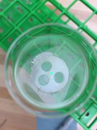 | 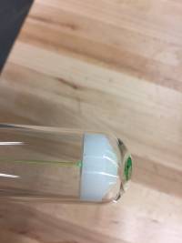 | 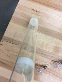 |
As discussed more later, this design did not work.
Procedure
The standard procedure recorded here was used, with minor modifications. A large batch of scintillator mixture was made in an Erlenmeyer flask (styrene with inhibitor removed, PPO, POPOP, and BPO) and sonicated for 10 minutes. Desired amounts of this scintillator mixture were measured into Pyrex 9800 test tubes. Dopants were added at this stage, unlike in previous trials where the fluors were added after the dopant.
| Figure 2: Setup for large scintillator batches |
|---|
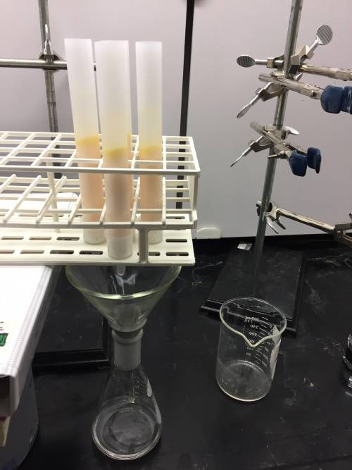 |
| 5 tubes with alumina were placed above a funnel, which was placed directly over an Erlenmeyer flask |
| List of Samples Fabricated (DMR-15) | Sample Description | Notes Before Polymerization | Notes During Polymerization | Notes After Polymerization |
|---|---|---|---|---|
| A | Unloaded, with fluors | Some air seems to have leaked out of balloon before being placed into the bath. Not sure as to the reason. | Sample A was the only sample to crack during the cooldown process. Crazing can be seen where the vial broke. | Bubble at very bottom of sample. Notable crazing in one spot on side of sample. |
| B | Unloaded, with fluors, with fiber in situ | Fiber came unattached from bottom holder. In future trials, fiber should be glued to mount before placing mount inside vial. | Vial B exploded 1 minute after being placed in bath. Shattering was caused by poor design of fiber holder. In future trials with WLS fiber, vial should be in small water bath and carefully watched for shattering soon after transfer to bath. | None. |
| C | 0.055% Gd, loaded with Gd(i-Pr)3 | Sample appeared slightly cloudy after filtration. | Bubbles at bottom of sample. Minor crazing along the sides that may polish off. Hard to tell sample quality prior to cutting and polishing. | |
| D | 0.055% Gd, loaded with Gd(TMHD)3 | Sample appears cloudy despite filtration. Minor crazing along the sides that may polish off. Hard to tell sample quality prior to cutting and polishing. | ||
| E | 0.1% Gd, loaded with Gd(i-Pr)3 | Undissolved compound equally suspended throughout styrene. | Undissolved isoproxide spread 2.5 cm from bottom of sample | |
| F | 0.1% Gd, loaded with Gd(TMHD)3 | All of compound appears to have dissolved. | Some white compound present in bottom of vial. | Bubbles at bottom of sample. Minor crazing along the sides that may polish off. Hard to tell sample quality prior to cutting and polishing. |
| G | 0.5% Gd, loaded with Gd(i-Pr)3 | Undissolved compound equally suspended throughout styrene. | Sample appeared to be fully polymerized before it was removed from vial. Upon breaking of the vial, it was discovered that the bottom half of the scintillator sample (appeared quite yellow) did not fully polymerize. The bottom half of the sample was still a viscous liquid. There was a clear separation between the undissolved isopropoxide and the scintillator about 5cm from the bottom of the test tube. | |
| H | 0.5% Gd, loaded with Gd(TMHD)3 | All of compound appears to have dissolved, styrene appears more yellow than sample A and sample F. | No undissolved compound. | Sample was completely unpolymerized when removed from the bath. The sample was more yellow than Sample A, but was in a viscous liquid state rather than a plastic. |
| Figure 3: Samples before being placed in water bath |
|---|
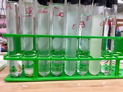 |
| From left to right: A, B, C, D, E, F, G, H |
Shortly (~1 min) after placing vials in water bath, DMR-15B shattered. A hole in the bottom fiber holder was not made to let styrene flow beneath the holder. While some did come through the WLS fiber hole, there was still air in this region. A hole should be added to the bottom holder before retrying in situ WLS fiber.
Conclusions, Notes for Future Runs
The samples should be bubbled in nitrogen for longer. 30-60 min was recommended for 20g samples but maybe 2 hrs should be used for 50g samples.
I am thoroughly perplexed in regards to the results I have observed. First, a new trial for the WLS fiber should be done. Second, it is unclear why sample D was cloudy even after filtration. Third,